Methodological Aspects of Advanced Laser Microtomy for plastic embedded tissue and materials
Dr. Emmanuel Loeb
Introduction
Plastic embedding was originally designed for bone histology, to avoid the decalcification process and later it was adjusted to solid, and implanted medical device research:
- Perfect method for rigid devices.
- Suitable for morphometric digital evaluation.
- Compatible with most staining procedures.
- Laser microtomy – advanced technique for delicate fine sections production.
- Relatively thin sections – up to 8-15 micron.
- Excellent histological image on a high-power field (magnification).
- High quality slides for documentation.
- Usually used for bone pathology, cardiac stents, tooth implants, orthopedic hard devices, regenerative medicine and tissue engineering (implants, scaffolds).
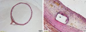

An example of a medical device implantation in a rabbit tissue with an inflammatory reaction.
The process:
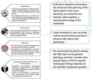

The advanced laser dissection: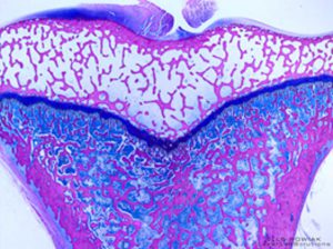

Laser microtomy can overcome fundamental limits of classical (hard tissue) microtomy and ground sectioning technology required for histological analysis in medical device and implant development:
Fast and easy cutting of undecalcified hard tissue and a broad range of implants and biomaterials
Semi-serial sectioning based on minimal material loss possible
Minimization of sectioning artefacts due to contact free cutting
Preservation of the implant-tissue interface
Quality control of sectioning via Optical Coherence Tomography.
Some nice examples:
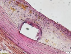
Coronary artery, stent with the increase of neointima. Note the details on a high magnification.

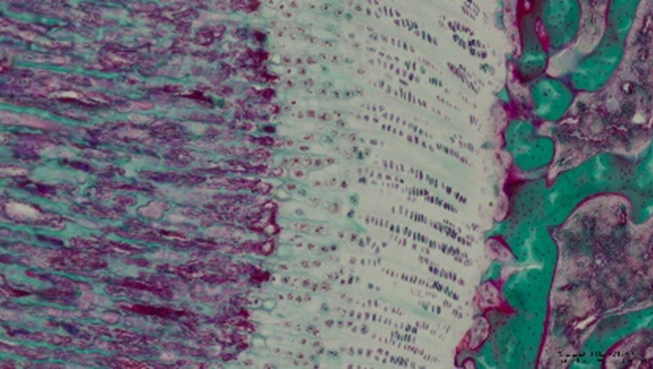
Rat knee, Masson Goldner
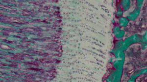

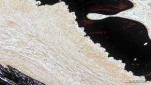
Polymer in bone, mineralization grade using the Von Kossa stain.
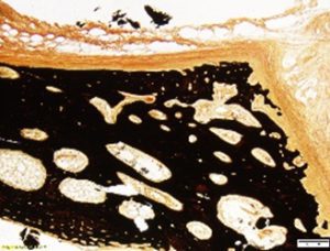

polymer implant, Von Kossa stain.
